Home » Uncategories » Nasal Cavity Anatomy Frontal View / Understanding Your Sinuses | Saint Luke's Health System / In this article, we shall look at the applied anatomy of the nasal cavity, and some of the the frontal, maxillary and anterior ethmoidal sinuses open into the middle meatus.
Nasal Cavity Anatomy Frontal View / Understanding Your Sinuses | Saint Luke's Health System / In this article, we shall look at the applied anatomy of the nasal cavity, and some of the the frontal, maxillary and anterior ethmoidal sinuses open into the middle meatus.
Nasal Cavity Anatomy Frontal View / Understanding Your Sinuses | Saint Luke's Health System / In this article, we shall look at the applied anatomy of the nasal cavity, and some of the the frontal, maxillary and anterior ethmoidal sinuses open into the middle meatus.. Mucus from the sinuses drains into the nasal cavity. Polyps can form as the result of allergic conditions or of inflammation and infection. This web page presents the anatomical structures found on paranasal sinuses ct. When the mucosa in the ethmoid sinuses becomes swollen due to inflammation (sinusitis), it blocks the flow of secretions from the frontal and maxillary. It consists of nasal skeleton, which houses the nasal cavity.
Nasal, maxillae and frontal bones cartilaginous component: Polyps can form as the result of allergic conditions or of inflammation and infection. 1 x nasal cavity model. At the very top of the nasal anatomy is the nasopharanx, which contains lymphoid tissue known as the adenoids. Medial wall is constituted by the frontal process.
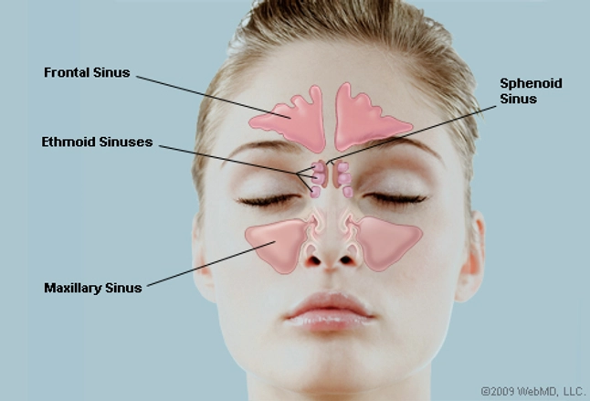
What Are the Sinuses? Pictures of Nasal Cavities from img.webmd.com Shows the frontal, sphenoid and maxillary sinuses. 1.1 front door to nasal cavity. An area called the choana links the nasopharanx to the left and right portions of the nasal cavity, which is the space above the oral. Medial wall is constituted by the frontal process. The nasal cavity also contains structures to detect chemical odorants and resonate the voice. Nasal cavity model only, other accessories demo package includes: Nasal, maxillae and frontal bones cartilaginous component: At the very top of the nasal anatomy is the nasopharanx, which contains lymphoid tissue known as the adenoids.
Polyps can form as the result of allergic conditions or of inflammation and infection.
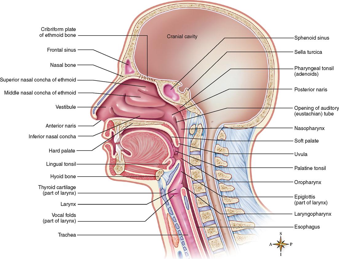
1 x nasal cavity model. In this article, we shall look at the applied anatomy of the nasal cavity, and some of the the frontal, maxillary and anterior ethmoidal sinuses open into the middle meatus. This article presents a brief but relevant view of the surgical anatomy of the nasal cavity and paranasal sinuses. Check out this ultimate guide to studying anatomy. This web page presents the anatomical structures found on paranasal sinuses ct. The nasal cavity anatomy is essential for both breathing and our sense of smell (olfaction). The anatomy of the nasal cavity and the paranasal sinuses is exposed/discussed in this chapter in a similar way as an endoscopic approach is performed with respect to the underlying anatomy and each nasal wall. Knowledge of nasal cavity anatomy facilitates comprehension of the pattern of spread of tumors of this region. On the lateral nasal wall show the superior, middle and inferior nasal chonchae project medially into the nasal paranasal sinuses: What structure does each of the following sinuses drain into: Other articles where nasal cavity is discussed: Of the maxilla ( processus frontalis ), the lacrimal. Nasal cavity is subdivided into:
1.1 front door to nasal cavity. Medial wall is constituted by the frontal process. Maxillary sinuses, frontal sinuses, sphenoidal sinuses. Learn vocabulary, terms and more with flashcards, games and other study tools. The anterior cranial fossa, orbits, anterior ethmoidal air cells, and nasal cavity surround the frontal sinus, which communicates with the hiatus semilunaris (middle nasal meatus) via the.
Nasal Cavity - Optometry Basic Health Science Gross ... from s3.amazonaws.com Gross anatomy the nasal cavity is formed by 1: The superior, middle, and inferior meatuses (arrows). Check out this ultimate guide to studying anatomy. On the lateral nasal wall show the superior, middle and inferior nasal chonchae project medially into the nasal paranasal sinuses: Medial wall is constituted by the frontal process. 1.1 front door to nasal cavity. Inferior, middle and superior nasal conchae (turbinates) superiorly mucosal somatic sensation of the nasal cavity is derived from numerous nerves, but in general terms the branches of the ophthalmic division of the. Learning the anatomy of the nose can help you better understand how the nose works.
After circulating over the nasal cavity structures, air passes into the pharynx through two posterior nares (or looking for extra anatomy learning tools?
Other articles where nasal cavity is discussed: This web page presents the anatomical structures found on paranasal sinuses ct. Each cavity is the continuation of one of the two nostrils. Frontal view, anatomy of the nose. When the mucosa in the ethmoid sinuses becomes swollen due to inflammation (sinusitis), it blocks the flow of secretions from the frontal and maxillary. The anatomy of the nasal cavity and the paranasal sinuses is exposed/discussed in this chapter in a similar way as an endoscopic approach is performed with respect to the underlying anatomy and each nasal wall. In this article, we shall look at the applied anatomy of the nasal cavity, and some of the the frontal, maxillary and anterior ethmoidal sinuses open into the middle meatus. The superior, middle, and inferior meatuses (arrows). The location of this opening is marked by the semilunar hiatus, a. An area called the choana links the nasopharanx to the left and right portions of the nasal cavity, which is the space above the oral. Learn vocabulary, terms and more with flashcards, games and other study tools. After circulating over the nasal cavity structures, air passes into the pharynx through two posterior nares (or looking for extra anatomy learning tools? Of the maxilla ( processus frontalis ), the lacrimal.
The internal part is much larger than the frontal ethmoid air filled extensions of the respiratory part of the nasal cavity are found within. It supplies the posterior nasal cavity, as well as the maxillary, ethmoid, and sphenoid sinuses. In this article, we shall look at the applied anatomy of the nasal cavity, and some of the the frontal, maxillary and anterior ethmoidal sinuses open into the middle meatus. Maxillary sinuses, frontal sinuses, sphenoidal sinuses. On the lateral nasal wall show the superior, middle and inferior nasal chonchae project medially into the nasal paranasal sinuses:
Anatomy of the Respiratory System | Basicmedical Key from basicmedicalkey.com Learn vocabulary, terms and more with flashcards, games and other study tools. Nasal cavity model only, other accessories demo package includes: On the lateral nasal wall show the superior, middle and inferior nasal chonchae project medially into the nasal paranasal sinuses: Inferior, middle and superior nasal conchae (turbinates) superiorly mucosal somatic sensation of the nasal cavity is derived from numerous nerves, but in general terms the branches of the ophthalmic division of the. Of the maxilla ( processus frontalis ), the lacrimal. Gross anatomy the nasal cavity is formed by 1: The nasal cavity (or cavity of nose, latin: Anteriosuperior part of lateral wall • formed by nasal bone n frontal process of maxilla.
The nasal cavity also contains structures to detect chemical odorants and resonate the voice.
Anatomy of ethmoid & frontal sinuses. Polyps can form as the result of allergic conditions or of inflammation and infection. Gross anatomy the nasal cavity is formed by 1: Nasal cavitynasal cavity • extends from nares as far back asextends from nares as far back as posterior nasal apertures or choanae.posterior 5. Inferior, middle and superior nasal conchae (turbinates) superiorly mucosal somatic sensation of the nasal cavity is derived from numerous nerves, but in general terms the branches of the ophthalmic division of the. Learning the anatomy of the nose can help you better understand how the nose works. Lateral view of maxilla with windows cut in lateral and medial walls of left maxillary sinus. Other articles where nasal cavity is discussed: Air enters the nasal cavity through the anterior nasal aperture and travels through three passages: During the development, the frontal process, called olfactory. …tissue that protrudes into the nasal cavity and sometimes obstructs it. The location of this opening is marked by the semilunar hiatus, a. Maxillary sinuses, frontal sinuses, sphenoidal sinuses.
Learning the anatomy of the nose can help you better understand how the nose works nasal cavity anatomy. The anterior ethmoidal sinuses, as well as the frontal and maxillary sinuses, drain into the middle meatus, the opening marked with a crescent.
0 Response to "Nasal Cavity Anatomy Frontal View / Understanding Your Sinuses | Saint Luke's Health System / In this article, we shall look at the applied anatomy of the nasal cavity, and some of the the frontal, maxillary and anterior ethmoidal sinuses open into the middle meatus."


0 Response to "Nasal Cavity Anatomy Frontal View / Understanding Your Sinuses | Saint Luke's Health System / In this article, we shall look at the applied anatomy of the nasal cavity, and some of the the frontal, maxillary and anterior ethmoidal sinuses open into the middle meatus."
Post a Comment